Body Imaging
Originally published in 1990
Body
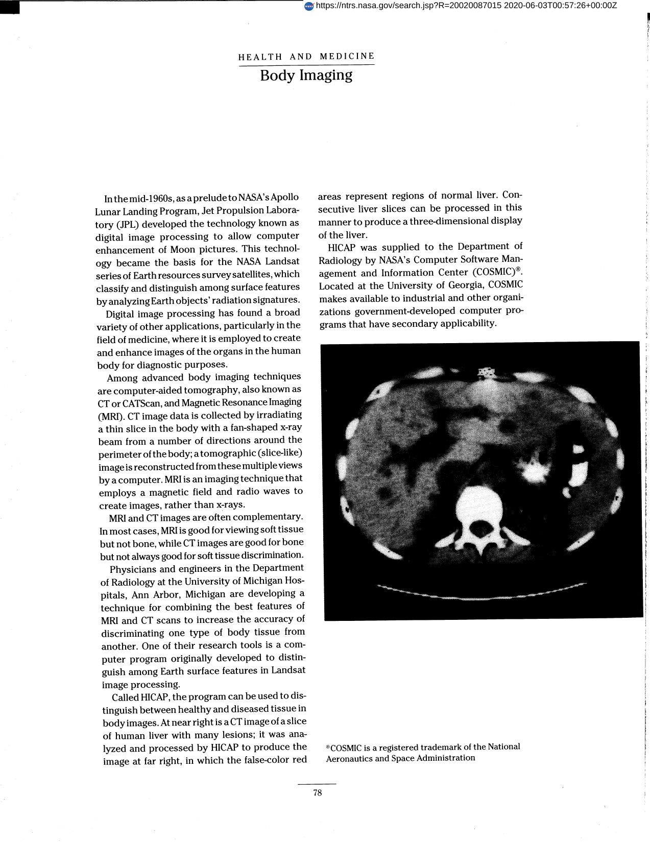
Magnetic Resonance Imaging (MRI) and Computer-aided Tomography (CT) images are often complementary. In most cases, MRI is good for viewing soft tissue but not bone, while CT images are good for bone but not always good for soft tissue discrimination. Physicians and engineers in the Department of Radiology at the University of Michigan Hospitals are developing a technique for combining the best features of MRI and CT scans to increase the accuracy of discriminating one type of body tissue from another. One of their research tools is a computer program called HICAP. The program can be used to distinguish between healthy and diseased tissue in body images.
Full article: http://hdl.handle.net/hdl:2060/20020087015
Abstract
Magnetic Resonance Imaging (MRI) and Computer-aided Tomography (CT) images are often complementary. In most cases, MRI is good for viewing soft tissue but not bone, while CT images are good for bone but not always good for soft tissue discrimination. Physicians and engineers in the Department of Radiology at the University of Michigan Hospitals are developing a technique for combining the best features of MRI and CT scans to increase the accuracy of discriminating one type of body tissue from another. One of their research tools is a computer program called HICAP. The program can be used to distinguish between healthy and diseased tissue in body images.

Body Imaging

Body Imaging

