Noninvasive Test Detects Cardiovascular Disease
Originating Technology/NASA Contribution
For decades now, NASA has been sending spacecraft throughout the galaxy. Once in the cosmos, these crafts use advanced cameras to create images of corners and crevices of our universe never before seen and then transmit these pictures back to laboratories on Earth, where government scientists then ask themselves: What exactly are we looking at?
That question is answered at NASA’s Jet Propulsion Laboratory (JPL) in the Image Processing Laboratory, founded in 1966 to receive and make sense of spacecraft imagery. There, NASA-invented VICAR (Video Image Communication and Retrieval) software has, through the years, laid the groundwork for understanding images of all kinds. The original software, created by a JPL team of three, Robert Nathan, Fred Billingsley, and Robert Selzer, is in use even to this day, although with greater accuracy and effectiveness due to decades of advancements.
The imaging division at JPL has grown increasingly sophisticated over the years, developing new processes and technologies to handle increasingly complex acquisitions from each NASA space mission, from the Voyager images of Saturn and Jupiter taken in the 1970s, to the new imagery captured by the Mars Reconnaissance Orbiter in late 2006 that suggests water still flows on Mars, opening the possibility that the Red Planet could perhaps support some forms of life.
Partnership
Selzer, from the original VICAR team, has made the NASA imaging technology his life’s work, spending 46 years as a NASA employee and continuing to work on its advancement even after his retirement from JPL. Selzer received many NASA awards for technical achievement, including the prestigious Technology and Application Program Exceptional Achievement Medal.
During the last 15 years of his career as a government scientist, he, as head of the JPL Biomedical Image Processing Laboratory, was working on using the imaging technology for health care diagnosis.
The project began when the imaging team developed the idea of using the VICAR software to analyze X-ray images of soft tissue. Typically, the X-ray is ineffective when used to analyze soft tissues, though the researchers were curious to see if the imaging software could broaden the application of this readily available diagnostic procedure. The results were interesting, but too much quality was lost in transferring the pictures into a digital format for analysis. Still, the idea seemed feasible, so, with several grants from NASA, the testing continued.
Selzer’s JPL team, partnering with scientists from the University of Southern California under the direction of the late Dr. David Blankenhorn and Dr. Howard Hodis, director of the Atherosclerosis Research Unit at the school’s Keck School of Medicine, began to image X-rays of arteries. With marginal success using X-rays, they came upon the idea of using the same methodology but applying it to ultrasound imagery, which was already digitally formatted. The new approach proved successful for assessing amounts of plaque buildup and arterial wall thickness, direct predictors of heart disease.
Testing continued, and the team, buoyed by its successes, began looking for outside funding and methods of distribution. At this point, Gary F. Thompson entered the picture.
Thompson has a history of heart disease in his family. The first male in many generations to live past age 50, and the last living male in his family line, Thompson comes from a long line of active, athletic men who, with no prior symptoms, suffered fatal heart-related events. A lifelong athlete who had boxed in the New York City Golden Gloves tournament in his youth and ran his first marathon in 1975, Thompson was understandably concerned, but feeling confident, when he approached his 50th birthday. He had the family history working against him, but he was also in prime shape. To celebrate his half-century mark, he planned to run three marathons: Los Angeles, New York, and Boston, and he underwent a battery of medical tests, all of which confirmed that he was in perfect health, without any signs of cardiovascular disease.
Seven days after his birthday, Thompson ran the Los Angeles marathon. At the 15th mile, he started experiencing back pain. By the 20th mile, it became so excruciating, that he stopped running and sought the help of a police officer who was monitoring the race. Later, at the hospital, doctors confirmed that he had suffered a moderate heart attack and lost 48 percent of his heart muscle. The modern medical testing, he realized, had failed him. Luckily for Thompson, compared to most men his age, for him to have 52 percent of his heart working was the equivalent of 127 percent, because he was so athletic.
Months later, at dinner with David Baltimore, then president of the California Institute of Technology (Caltech), Thompson asked whether there were any new heart-related breakthroughs from the esteemed university and was surprised to hear that there actually was, indeed, a new technology, but that it had been developed at JPL. It was a noninvasive diagnostic system with the ability to accurately predict heart health. Baltimore offered to set up an appointment for Thompson at the University of Southern California hospital where this new method was being tested, but Thompson declined the privileged treatment.
Instead, he went in on his own, unannounced, and without revealing his family history or recent heart attack. Thompson, who, despite the recent event, gave every impression of great health, met with a technician, who asked him to loosen his collar and then performed an ultrasound scan on either side of his neck, the location of the carotid arteries. When the results were in, the technician told Thompson that he needed to meet with the doctor immediately. The test showed that Thompson had the thickest artery walls of the over 3,000 other people tested, a direct indicator that he was in danger of a heart attack or stroke.
Thompson was impressed. This new device had managed to do what all of the other tests had failed to do: give him an accurate reading of his heart health. Thompson, a hard-charging entrepreneur, met with the researchers Selzer and Hodis and told them that they needed to get this technology into the hands of physicians. They agreed. Thompson developed a business plan, secured an exclusive license for the JPL-developed technology from Caltech, and invested his own money to start Medical Technologies International Inc. (MTI), located in Palm Desert, California. Selzer, after retiring from JPL, joined in and now serves as the company’s chief engineer.
Product Outcome
MTI licensed 14 research institutions around the world for pre-U.S. Food and Drug Administration (FDA) clearance, research-only use of the analysis software and then incorporated feedback from these groups into the new clinical product it was developing. It patented the new developments and then submitted the technology to a rigorous review process at the FDA, which cleared the device for public use. MTI also filed with the American Medical Association to have the device given a dedicated Current Procedural Technology (CPT) code for insurance purposes, thus encouraging more doctors to offer this test to patients.
The patented software is being used in MTI’s ArterioVision, a carotid intima-media thickness (CIMT) test that uses ultrasound image-capturing and analysis software to noninvasively identify the risk for the major cause of heart attack and strokes: atherosclerosis.
The term atherosclerosis comes from Greek, with athero meaning a gruel or paste (plaque) and sclerosis meaning hardness. It is just that, a buildup of cholesterol and fatty substances in the arteries, combined with arterial hardening. The result is that blood flow through the heart is restricted, hampering oxygen supply. Heart attacks occur when the heart does not get the necessary oxygen, and strokes are the result of oxygen not reaching the brain. Atherosclerosis, referred to by the American Heart Association (AHA) as the “silent killer,” initially has no discernable symptoms until one or more of the arteries becomes so congested that these major, sometimes fatal, problems occur. The AHA estimates that two out of three unexpected cardiac deaths occur without prior symptoms.
In fact, astronaut Edward White, the first astronaut to ever perform a space walk, and one of the three space pioneers to die during the Apollo launch pad tragedy in 1967, was thought by most to be in perfect health, having successfully passed all of the rigorous astronaut testing. An autopsy directly after the accident, though, revealed that he had extreme thickening of the arteries and showed most signs of heart disease. With this particular disease, though, as has been mentioned previously, often the only symptoms are either heart attack or stroke.
ArterioVision provides a direct measurement of atherosclerosis before it causes these “symptoms” by safely and painlessly measuring the thickness of the first two layers of the carotid artery wall using an ultrasound procedure and advanced image-analysis software. The carotid artery, located on both sides of the neck, is the largest artery that is near the surface of the skin. It supplies blood to the brain, and it can therefore be examined noninvasively. ArterioVision essentially performs a skin surface imaging “biopsy” to examine the arterial wall.
As evidenced by the battery of tests Thompson underwent before his heart attack, diagnostic tools for atherosclerosis screening are far from advanced. Atherosclerosis begins in the abdomen and ascends to the heart and the carotid arteries. A diagnostic tool for examining it in the abdomen is the computerized tomography (CT) scan, but CT comes with a certain level of risk and at a great financial cost. Unlike ultrasound, which is safe enough that it is used on unborn babies, CT scans rely on radiation to produce results. While this procedure is still new enough that the risks are relatively unknown, the National Toxicology Program, a division of the U.S. Department of Health and Human Services, has recently declared X-rays carcinogenic. Similarly, researchers at Columbia University have estimated that radiation for a full-body scan is roughly equivalent to the same amount of radiation exposure experienced by people within 1.5 miles of the atomic bombs dropped on Nagasaki and Hiroshima. In other terms, it is roughly equivalent to the radiation from 200 chest X-rays. As an added disadvantage, X-ray machines and the production of radiation cost a great deal of money.
The NASA-based technology has none of these problems. It poses no risks, and it is relatively inexpensive. The imaging technology can distinguish between 256 different shades of gray and differentiate nuances at a sub-pixel level of interpolation, making it the most accurate in this field, and it is compatible with all existing ultrasound machines, making it more readily accessible to physicians.
While ArterioVision is not the only FDA-cleared CIMT tool on the market, it is the only one that offers a predictive report for the physician and patient. It explains the significance of test results using a proprietary database and JPL-developed algorithms, and can extrapolate to show percentile of risk.
One particular feature of the report is the revelation of arterial age. It can show the patient that while he may be 50 years old, his arteries may be the equivalent of patients 75 years old. This real-world number is something patients can identify with and helps promote compliance with drug therapies and other forms of treatment—one of the most difficult aspects of preventing and treating heart disease. Physicians often lament that they stress the importance of lifestyle changes to their patients, but since heart disease does not initially come with symptoms, but instead with potentially fatal events, it is often difficult to impress upon the patients the urgency of taking care of their hearts. The ArterioVision patient report provides a significant warning sign and gives concrete examples.
The report then becomes part of a serial examination. Atherosclerosis can be reversed with strategies like exercise, diet change, weight loss, smoking cessation, and cholesterol-lowering medication; and the patient can see, after implementing a new strategy, if that one is working.
Currently, the technology is in all 50 states, and in many countries throughout the world. MTI is continuing to push this life-saving technology and is rapidly expanding its sales force in an effort to live up to its company credo, “Making a Positive Difference Every Day.”
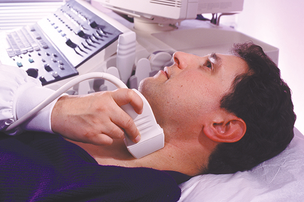
ArterioVision is a CIMT test that uses ultrasound image-capturing and analysis software to noninvasively identify the risk for the major cause of heart attack and strokes: atherosclerosis.
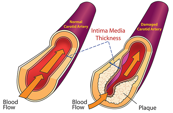
Atherosclerosis is a condition in which fatty material collects along the walls of arteries. This fatty material thickens, hardens, and may eventually block the arteries.
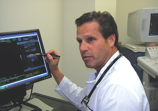
ArterioVision is the only CIMT tool on the market that offers a predictive report for physicians and patients. It explains the significance of test results using a proprietary database and JPL-developed algorithms and can extrapolate to show percentile of risk.

Images from the Mars Reconnaissance Orbiter suggest that liquid water has flowed on Mars within the past 6 years. These pictures were analyzed and clarified with imaging software technologies from NASA’s Jet Propulsion Laboratory.
